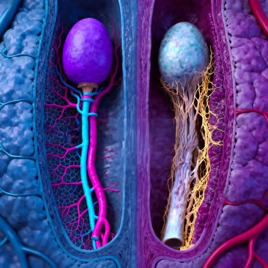
The human body is made up of many complex systems that work together to maintain life and allow the body to grow, heal, and reproduce. Among these systems, the reproductive system plays a vital role in ensuring the continuation of human life. The image above appears to represent a detailed illustration of structures within the male reproductive system, specifically focusing on the testes and spermatic pathways, showing how biological structures are connected through networks of blood vessels and ducts.
Although illustrations like this are often stylized to help people visualize anatomy more clearly, they can provide a helpful starting point for understanding how the reproductive organs function and how different biological components interact.
This article explores the anatomy of the male reproductive system, focusing on the testes, the transport of sperm, and the networks of blood vessels and nerves that support reproductive health.
The Role of the Male Reproductive System
The male reproductive system has two primary biological functions:
- Producing sperm cells, which carry genetic material.
- Producing hormones, particularly testosterone, which plays a major role in male development and health.
These functions involve several organs that work together as a coordinated system. Key parts include:
- Testes
- Epididymis
- Vas deferens
- Seminal vesicles
- Prostate gland
- Penis
Each of these components contributes to the production, maturation, and delivery of sperm.
The image shown appears to focus on the internal structures connected to the testes, highlighting how sperm cells travel through small ducts while being supported by surrounding blood vessels and tissues.
The Testes: The Starting Point of Sperm Production
The testes, often referred to as testicles, are two oval-shaped organs located inside the scrotum. Their primary role is to produce sperm and the hormone testosterone.
Inside each testis are tiny coiled tubes called seminiferous tubules. These microscopic structures are where sperm cells begin their development. Millions of sperm cells are produced daily within these tubules.
The image likely represents this internal process, with the upper round structure symbolizing the testicle and the lower connecting pathways illustrating how sperm travel outward from the testes.
The Process of Spermatogenesis
The formation of sperm cells is called spermatogenesis, a complex biological process that takes place within the seminiferous tubules.
This process occurs in several stages:
- Stem cells divide to create early sperm cells.
- These cells gradually develop a head and tail.
- They mature into fully formed sperm capable of movement.
The entire process takes approximately 64 to 74 days in humans.
After sperm cells form inside the testes, they are not yet fully mature. They must travel to another structure called the epididymis to gain the ability to swim and fertilize an egg.
The Epididymis: Where Sperm Mature
The epididymis is a long, tightly coiled tube that sits along the back of each testis. Though small in size, it plays a crucial role in sperm development.
Once sperm leave the seminiferous tubules, they move into the epididymis where they:
- Mature and gain mobility
- Become capable of fertilization
- Are stored until ejaculation
The structure shown in the illustration may represent this connection between the testicle and the narrow duct that carries sperm away from it.
The epididymis is highly specialized, with microscopic channels that allow sperm to move gradually while being nourished by surrounding cells.
The Vas Deferens: The Transport Highway
Once sperm have matured in the epididymis, they enter the vas deferens, a muscular tube that transports them further through the reproductive system.
The vas deferens connects the epididymis to other reproductive structures deeper in the pelvis.
When ejaculation occurs, muscles surrounding the vas deferens contract rhythmically, pushing sperm forward. These contractions allow sperm to travel quickly toward the urethra, where they mix with fluids produced by other glands.
The elongated tube shown in the image likely represents this transport pathway.
Blood Vessels and Nerve Networks
The illustration also appears to highlight a network of blood vessels and nerves surrounding the reproductive structures.
These networks are essential for several reasons:
Blood Supply
Blood vessels deliver oxygen and nutrients to reproductive tissues. They also help regulate temperature, which is critical for sperm production.
Sperm cells develop best at temperatures slightly lower than normal body temperature. This is one reason the testes are located in the scrotum outside the main body cavity.
The pampiniform plexus, a network of veins surrounding the testicular artery, helps cool incoming blood and maintain an ideal temperature for sperm production.
Nerve Signals
Nerves play an important role in reproductive function. They help control:
- Muscle contractions during ejaculation
- Sensory responses
- Hormonal signaling
Without these nerve pathways, many reproductive processes would not function properly.
Hormones and Reproductive Health
The testes are not only responsible for producing sperm but also for releasing hormones. The most important of these hormones is testosterone.
Testosterone influences many aspects of male health, including:
- Development of male physical characteristics
- Muscle and bone growth
- Libido and sexual function
- Mood and energy levels
Hormones are regulated through communication between the testes and the brain, particularly the hypothalamus and pituitary gland.
These organs release signals that tell the testes when to produce testosterone and sperm.
Why Medical Illustrations Matter
Images like the one shown can be helpful educational tools. Medical illustrations often exaggerate colors or shapes to make anatomical structures easier to distinguish.
For example:
- Blood vessels may be shown in bright colors.
- Nerve networks may appear highlighted.
- Tissue layers may be separated for clarity.
These artistic choices help students and readers understand complex anatomy more easily.
However, the actual structures inside the body are much smaller and more compact than they appear in illustrations.
Maintaining Reproductive Health
Just like any other system in the body, the male reproductive system requires proper care to remain healthy.
Several lifestyle factors can influence reproductive health, including:
Nutrition
A balanced diet containing vitamins and minerals supports hormone production and overall health.
Important nutrients include:
- Zinc
- Vitamin D
- Omega-3 fatty acids
- Antioxidants
Physical Activity
Regular exercise helps regulate hormones and maintain healthy blood circulation.
However, excessive stress or overtraining can sometimes disrupt hormonal balance.
Sleep
Quality sleep is essential for hormone regulation. Testosterone levels tend to peak during sleep cycles.
Chronic sleep deprivation may lead to reduced hormone production over time.
Environmental and Lifestyle Factors
Several environmental factors may influence reproductive health.
These can include:
- Exposure to certain chemicals
- Excessive heat
- Smoking
- High levels of stress
Maintaining a healthy lifestyle and minimizing harmful exposures can support long-term reproductive function.
Medical Conditions That Affect the Testes
There are several medical conditions that can affect the structures shown in the illustration.
Some of the most common include:
Varicocele
A varicocele occurs when veins in the scrotum become enlarged. This condition can affect blood flow and sometimes influence sperm production.
Epididymitis
This condition involves inflammation of the epididymis, often caused by infection.
Symptoms may include swelling, pain, and discomfort.
Testicular Torsion
Testicular torsion occurs when the spermatic cord twists, cutting off blood supply to the testis. It is considered a medical emergency and requires immediate treatment.
The Importance of Early Detection
Regular health checkups and awareness of bodily changes can help detect problems early.
Doctors often recommend that individuals become familiar with their own bodies so they can recognize unusual changes.
Early detection of medical issues can lead to better treatment outcomes.
Advances in Medical Research
Modern medical research continues to expand our understanding of reproductive health.
Scientists are studying:
- The genetics of fertility
- New treatments for reproductive disorders
- Improved surgical techniques
- Hormonal therapies
These advancements may help improve diagnosis and treatment options in the future.
Educational Value of Anatomical Models
Images like the one shown are frequently used in:
- Medical textbooks
- Educational websites
- Biology classrooms
- Health awareness campaigns
They allow students and readers to explore anatomy in ways that would otherwise be impossible without advanced medical equipment.
Three-dimensional illustrations can make it easier to visualize how organs and tissues connect and interact.
A Complex and Remarkable System
The male reproductive system represents a remarkable example of biological coordination. Multiple organs, tissues, and chemical signals work together to support the processes of hormone production and sperm development.
Although these processes happen largely without conscious awareness, they depend on delicate biological balances.
The structures depicted in the illustration—testes, ducts, blood vessels, and surrounding tissues—highlight the complexity of human anatomy.
Final Thoughts
Understanding the body’s anatomy is an important step toward appreciating how biological systems function and how they can be protected through healthy habits and medical care.
The image presented illustrates key components of the male reproductive system and the pathways through which sperm develop and travel. While stylized for educational purposes, it reflects real biological structures that play vital roles in reproduction and hormonal balance.
By learning more about these systems, individuals can make informed decisions about their health, recognize potential concerns earlier, and seek appropriate medical advice when needed.
The human body remains one of the most fascinating and intricate systems known, and studying its anatomy continues to reveal new insights into how life functions at its most fundamental levels.